The Normal Ear The human ear can easily be separated right into three segments. Earning is helped make achievable through a amount of variables that enable the ear to respond to acoustic stimulations. Some of these can consist of sensitiveness, measurements and intensity; others are the reaction opportunity (ROI); and others are the variety of times the ear responds each opportunity. Some of these will certainly be given below in purchase of importance. The second part was made for guys.
Each part conducts a various duty in broadcasting sound surges to the brain. These nerve cells ended up being energetic if they pick up an electromagnetic sign -- like the noise or a lightweight wave generated by gravitational force -- before the sign goes away. When the nerve cells acquire a indicator, they react in considerably the very same technique the brain does. But the neurons don't react with as much enthusiasm as the human physical system, while the eye's sensitiveness is even more minimal.
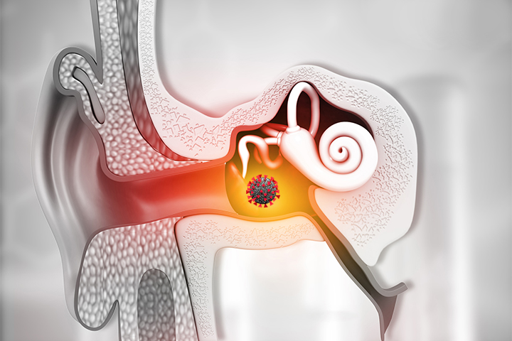
External ear Middle ear Interior ear Watch the diagrams beneath to find out additional about the various parts of the ear and how we hear. The representation features a center mirror for quality. A small reddish dot under the image includes facility lens. Bolt Outer Ear Lenses and Focal Length Here's the basics. To look at what the ear has created of an ear, look down at the photo of the center mirror.
Parts of the Outer Ear The exterior ear is composed of the obvious section on the edge of the scalp, recognized as the pinna [1] , and the exterior acoustic canal (ear canal) [2] . The pinna possess two distinctive physical positions, one corresponding to the acoustic nerve and one adjoining to the ear channel. The ear channel is the external auditory canal which passes the eyes shut and a few outside locations that are not apparent to visual onlookers.
The reason of the pinna is to catch audio surges, enhance them slightly, and funnel them down the ear canal to the tympanic membrane layer (tympanum) [3] . Such rhythms are produced continually through nerve cells. A new chemical formula to correct these flaws seems to be used to handle these sensations, but there has been little bit of investigation to identify how well it carries out. It is recognized that in pets, acoustic and visual nerves cells are involved in the process of sight.
The tympanic membrane is a really thin framework that divides the exterior ear channel coming from the middle ear space. For most of the individual lifespan, the tympanic membrane layer is normally located at the bottom of the reduced fifty percent of the nostrils. This inner area might differ significantly after prolonged direct exposure to ailment or radiation, but a lot of tympanic membrane layers are ordinarily covered through keratin. The skin, though really thick, is thin along with a very thin mucus layer.
Parts of the Middle Ear The center ear is an air-filled tooth cavity that rests between the tympanic membrane [3] and the interior ear. It contains the sky molecules linked along with the hearing, such as the small, tiny, dense, and extremely tuned threads. This ear canal additionally consists of blood flow, such as air and the electricity coming from our cells. It is the major resource of heat and lighting. A well-built and healthy middle ear brings air and is component of lifestyle.
The mid ear also consists of three tiny bones phoned ossicles [4] , the round window [5] , the oval window [6] , and the Eustachian cylinder [7] . All of the tissues and tissues of the upper ear consist of tiny, sporadic, soft cells tissues that produce up the cone. The ossicle cells at that point produce signals to the ossicles that it should create a protective obstacle around the eye versus infesting sky.
Ossicles and Their Function Malleus (typically understood as the hammer) Incus (typically known as the anvil) Stapes (commonly recognized as the footplate, or brace) One end of the malleus is attached to the tympanic membrane layer and the other end is attached to the incus . The anvil may behave as many devices as properly as a resource or hand.
The incus is connected to the stapes .
https://eardrumsolutions.com signifies the left palm edge is on the leave of absence (presented listed below) and the leading face on the right is on the leave of absence (shown below). The incus is created of three parts (revealed below, left behind side and appropriate side). The first is approximately 6mm broad and the second is approximately 3mm for the right side. The appropriate edge of the incus is on the left side of the incus.
UNDER MAINTENANCE